
Adult Health

Anatomy of the Heart
Heart Structure
- The heart is a muscular organ located in the mediastinum, encased in a thin sac called the pericardium.
Layers of the Heart
- Epicardium: The outermost layer, also known as the visceral pericardium.
- Myocardium: The middle layer responsible for the pumping action of the heart.
- Endocardium: The innermost layer lining the chambers and valves of the heart.
Heart Chambers and Valves
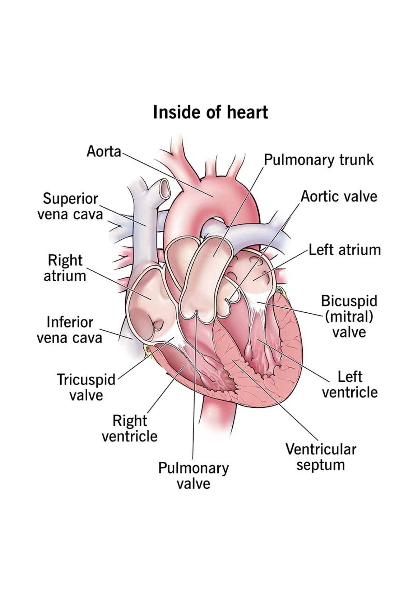
- The heart has four chambers: two atria (upper) and two ventricles (lower).
- Valves ensure unidirectional blood flow:
- Tricuspid Valve: Between the right atrium and right ventricle.
- Pulmonary Valve: Between the right ventricle and pulmonary artery.
- Mitral Valve: Between the left atrium and left ventricle.
- Aortic Valve: Between the left ventricle and aorta.
Blood Flow Through the Heart
Steps of Blood Flow
- Deoxygenated blood enters the right atrium via the superior and inferior vena cava.
- Blood flows through the tricuspid valve into the right ventricle.
- From the right ventricle, blood is pumped through the pulmonary valve into the pulmonary artery and to the lungs for oxygenation.
- Oxygenated blood returns to the left atrium via the pulmonary veins.
- Blood flows through the mitral valve into the left ventricle.
- The left ventricle pumps oxygenated blood through the aortic valve into the aorta, delivering it to the rest of the body.

Electrical Conduction System
Key Components
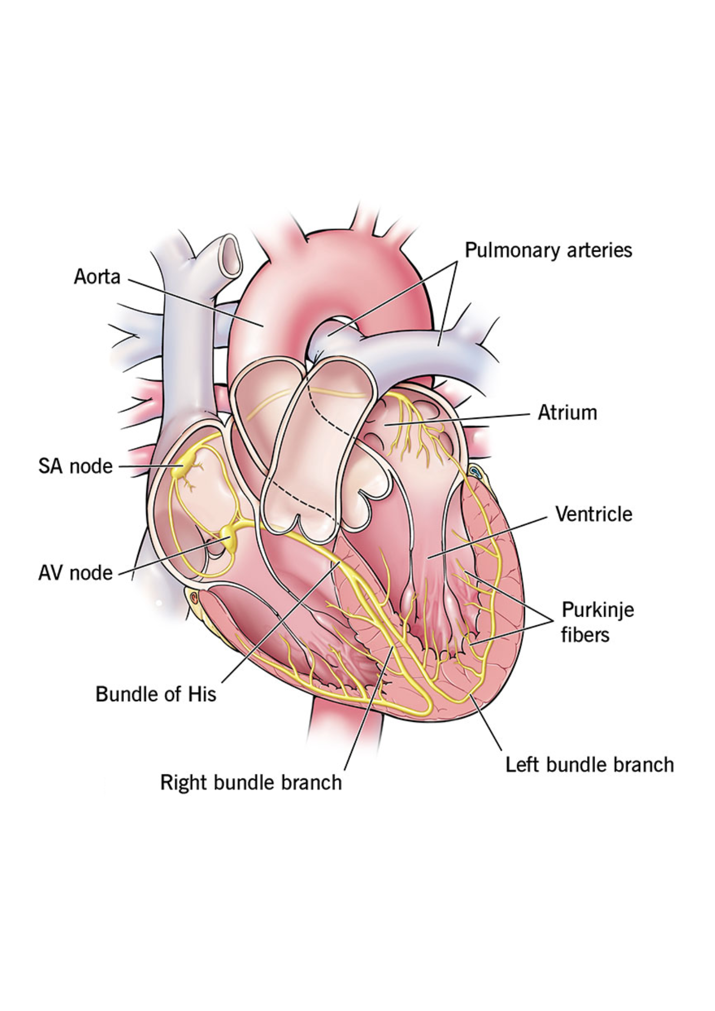
- Sinoatrial (SA) Node: The primary pacemaker, generating impulses at 60-100 bpm.
- Atrioventricular (AV) Node: Delays the impulse to allow ventricular filling, generating impulses at 40-60 bpm if needed.
- Bundle of His: Transmits impulses to the right and left bundle branches.
- Purkinje Fibers: Conduct impulses throughout the ventricles, generating contractions at 20-40 bpm if other pacemakers fail.
Hemodynamic Terms and Parameters
Hemodynamic Parameters
- Central Venous Pressure (CVP): Reflects right atrial pressure and preload.
- Normal Range: 2-6 mmHg.
- Clinical Relevance: Increased in fluid overload and heart failure; decreased in hypovolemia.
- Pulmonary Artery Wedge Pressure (PAWP): Estimates left atrial pressure and left ventricular preload.
- Normal Range: 6-12 mmHg.
- Clinical Relevance: Elevated in left-sided heart failure or mitral valve disorders.
- Mean Arterial Pressure (MAP): Average pressure in the arteries during one cardiac cycle.
- Formula: MAP = (Systolic BP + 2 × Diastolic BP) / 3.
- Normal Range: 70-100 mmHg.
- Clinical Relevance: Must be ≥60 mmHg to adequately perfuse vital organs.
- Systemic Vascular Resistance (SVR): Resistance faced by the left ventricle during systole.
- Normal Range: 800-1200 dynes/sec/cm5.
- Clinical Relevance: Increased in vasoconstriction and hypertension; decreased in septic shock.
- Stroke Volume (SV): Amount of blood ejected with each heartbeat.
- Normal Range: 60-120 mL/beat.
- Clinical Relevance: Affected by preload, afterload, and myocardial contractility.
Lab Values Related to the Cardiac System
- Troponin I:
- Definition: A highly specific marker for myocardial infarction (MI).
- Normal Range: Less than 0.03 ng/mL.
- Clinical Relevance: Elevates within 4-6 hours of MI and remains elevated for 7-10 days.
- BNP (B-Type Natriuretic Peptide):
- Definition: Released when the heart senses increased stretch or pressure.
- Normal Range: Less than 100 pg/mL.
- Clinical Relevance: Elevated in heart failure; higher levels indicate worse severity.
- CK-MB (Creatine Kinase-MB):
- Definition: An enzyme found in the heart muscle, elevated after myocardial injury.
- Normal Range: 0-5 ng/mL.
- Clinical Relevance: Rises within 3-6 hours and returns to normal within 2-3 days.
- Myoglobin:
- Definition: An oxygen-binding protein found in muscle, released during injury.
- Normal Range: 0-85 ng/mL.
- Clinical Relevance: Elevates early (within 2 hours) but is not cardiac-specific.
- Serum Potassium (K+):
- Normal Range: 3.5-5.0 mEq/L.
- Clinical Relevance: Hyperkalemia or hypokalemia can lead to life-threatening arrhythmias.
Cardiac Terms
- Preload: The volume of blood in the ventricles at the end of diastole.
- Afterload: The resistance the heart must overcome to eject blood during systole.
- Cardiac Output (CO): The amount of blood pumped by the heart per minute.
- Cardiac Index (CI): Cardiac output adjusted for body surface area.
- Stroke Volume (SV): The volume of blood ejected with each contraction.
- Ejection Fraction (EF): The percentage of blood ejected from the left ventricle during systole.
- Systemic Vascular Resistance (SVR): Resistance to blood flow in systemic circulation.
Cardiac Auscultation
Key Auscultation Areas
- Aortic Area: Located at the second intercostal space, right sternal border.
- Associated Murmurs: Systolic murmurs, such as aortic stenosis or aortic sclerosis.
- Pulmonic Area: Located at the second intercostal space, left sternal border.
- Associated Murmurs: Systolic ejection murmurs, such as pulmonic stenosis or atrial septal defect.
- Erb’s Point: Located at the third intercostal space, left sternal border.
- Associated Murmurs: Diastolic murmurs, such as aortic regurgitation or pulmonic regurgitation.
- Tricuspid Area: Located at the fourth intercostal space, left sternal border.
- Associated Murmurs: Holosystolic murmurs, such as tricuspid regurgitation or ventricular septal defect.
- Mitral Area (Apex): Located at the fifth intercostal space, midclavicular line.
- Associated Murmurs: Holosystolic murmurs (mitral regurgitation) and diastolic murmurs (mitral stenosis).
Clinical Notes
- Diastolic Murmurs: Always pathological and require further investigation.
- Systolic Murmurs: Can be physiological or pathological, depending on clinical context.
- Erb’s Point: Ideal for listening to both S1 and S2 sounds clearly.

Vital Signs and Diagnostics
Blood Pressure (BP)
- Normal Range: 120/80 mmHg.
- Hypertension: BP ≥ 140/90 mmHg, associated with increased risk of stroke, heart disease, and kidney damage.
- Hypotension: BP ≤ 90/60 mmHg, may cause dizziness, fainting, or shock.
- Clinical Tip: Ensure the correct cuff size and patient positioning for accurate readings.
Heart Sounds
- Normal Sounds: S1 (lub) and S2 (dub) indicate the closure of heart valves.
- Abnormal Sounds: Murmurs (graded I to VI), rubs, and gallops (e.g., S3 and S4).
- Clinical Tip: Auscultate using both the diaphragm and bell of the stethoscope to detect high- and low-frequency sounds.
Electrocardiogram (ECG/EKG) Intervals
- P Wave: Represents atrial depolarization. Duration: ≤ 0.12 seconds.
- PR Interval: Time between atrial depolarization and ventricular depolarization. Normal range: 0.12-0.20 seconds.
- QRS Complex: Represents ventricular depolarization. Duration: ≤ 0.12 seconds.
- QT Interval: Time from ventricular depolarization to repolarization. Duration: 0.36-0.44 seconds.
- ST Segment: Represents the time between ventricular depolarization and repolarization. Should be isoelectric (flat).
- T Wave: Represents ventricular repolarization. Inversion may indicate ischemia.
Clinical Diagnostics
- Chest X-Ray: Evaluates heart size, pulmonary congestion, and pleural effusion.
- Echocardiogram: Assesses heart structure, wall motion, and ejection fraction (EF).
- Stress Test: Monitors cardiac response to physical or pharmacologic stress.
- Holter Monitor: Continuous ECG monitoring over 24-48 hours to detect arrhythmias.
Tips for Accurate Assessment
- Ensure the patient is calm and seated for at least 5 minutes before measuring blood pressure.
- Auscultate heart sounds in all four key areas: aortic, pulmonic, tricuspid, and mitral.
- Verify ECG lead placement to avoid incorrect readings.
Introduction to Cardiac Arrhythmias
Cardiac arrhythmias are abnormalities in the electrical activity of the heart, leading to irregular heartbeats. These may be too fast, too slow, or erratic. Arrhythmias can originate in different areas of the heart and may significantly impact cardiac output and overall health.
Classification of Arrhythmias
Arrhythmias are classified based on their origin (atria or ventricles) and the type of irregularity. They are broadly categorized into:
- Bradyarrhythmias: Slow heart rhythms, such as sinus bradycardia.
- Tachyarrhythmias: Fast heart rhythms, such as sinus tachycardia or ventricular tachycardia.
- Fibrillation: Disorganized electrical activity, such as atrial fibrillation or ventricular fibrillation.
- Asystole: Complete absence of electrical activity in the heart.
Importance of Recognizing Arrhythmias
Early recognition and treatment of arrhythmias are crucial to prevent complications such as stroke, heart failure, or sudden cardiac death. Understanding the characteristics, causes, and management of each arrhythmia is essential for healthcare providers to deliver timely and effective care.
Common Symptoms of Arrhythmias
- Palpitations (feeling of rapid, fluttering, or pounding heartbeat).
- Shortness of breath or dyspnea.
- Fatigue or weakness.
- Lightheadedness or dizziness.
- Chest pain or discomfort.
- Syncope (fainting) or near-fainting episodes.
Diagnostic Tools
Diagnosing arrhythmias involves using various tools to evaluate heart rhythm and electrical activity. Common diagnostic methods include:
- Electrocardiogram (ECG/EKG): The primary tool for detecting and classifying arrhythmias.
- Holter Monitoring: Continuous ECG monitoring over 24-48 hours.
- Event Monitors: Used for intermittent arrhythmias not captured by standard ECG.
- Electrophysiology Studies (EPS): Invasive testing to identify arrhythmia triggers and guide treatment.

Sinus Bradycardia
Characteristics
- Rate: Less than 60 beats per minute.
- Characteristics:
- P Waves precede each QRS complex.
- PR interval is normal.
- QRS complex is normal.
- Rhythm is regular.
Causes
- Drugs (e.g., beta blockers).
- Vagal stimulation.
- Hypothermia.
- Sinus node involvement in MI.
- Can be normal in athletes.
Clinical Manifestations
- Often asymptomatic.
- Symptoms may include syncope, fatigue, and dizziness.
Management
- Treat the underlying cause.
- Administer atropine sulfate as prescribed.

Sinus Tachycardia
Characteristics
- Rate: 100-180 beats per minute.
- Other Features:
- P Waves precede each QRS complex.
- PR interval and QRS complex are normal.
- Rhythm is regular.
Causes
- Exercise or emotional stress.
- Fever or infection.
- Anemia, hypovolemia, or shock.
- Drugs (e.g., caffeine, nicotine).
Clinical Manifestations
- Often asymptomatic.
- May include palpitations, mild dizziness, or fatigue.
Management
- Treat the primary cause (e.g., infection, dehydration).
- Carotid sinus massage or beta blockers may reduce heart rate.
- Educate patients on avoiding stimulants (e.g., caffeine).


Atrial Fibrillation
Characteristics
- Atrial Rate: 350-600 bpm.
- Ventricular Rate: 120-200 bpm (if uncontrolled).
- Other Features:
- P wave is not discernible; irregular baseline.
- PR interval is not measurable.
- QRS complex is normal.
- Rhythm is irregularly irregular.
Causes
- Hypertension or coronary artery disease.
- Heart failure.
- COPD or thyrotoxicosis.
- Post-cardiac surgery.
Clinical Manifestations
- Palpitations or irregular pulse.
- Dyspnea or fatigue.
- Increased risk of thromboembolism (e.g., stroke).
Management
- Control ventricular rate using beta blockers or calcium channel blockers.
- Anticoagulation to prevent thromboembolism (e.g., warfarin, DOACs).
- Rhythm control with cardioversion or antiarrhythmic drugs.

Ventricular Tachycardia
Characteristics
- Rate: 100-250 beats per minute.
- Other Features:
- P wave is blurred in the QRS complex; no association with P wave.
- PR interval is not present.
- QRS complex is wide and bizarre; T wave is in the opposite direction.
- Rhythm is usually regular but may start and stop suddenly.
Causes
- Myocardial infarction (MI).
- Coronary artery disease (CAD).
- Hypokalemia or hyperkalemia.
- Mitral valve prolapse.
- Rheumatic heart disease or aneurysm.
- Pulmonary embolism.
- Anxiety or stress.
Clinical Manifestations
- Lightheadedness or dizziness.
- Weakness or fatigue.
- Dyspnea (difficulty breathing).
- Loss of consciousness in severe cases.
Management
Pulseless Ventricular Tachycardia
- Initiate cardiopulmonary resuscitation (CPR).
- Defibrillation as per ACLS protocol.
- Administer epinephrine or vasopressin.
- Consider advanced airway management (e.g., ET intubation).
Ventricular Tachycardia with Pulse
- If hemodynamically stable:
- Administer amiodarone or procainamide as per ACLS protocol.
- If hemodynamically unstable:
- Perform synchronized cardioversion.

Ventricular Fibrillation
Characteristics
- Rate: Rapid and uncoordinated; ventricles quiver instead of contracting effectively.
- Other Features:
- Rhythm is chaotic and irregular.
- QRS complexes are wide and irregular.
- P wave is not seen, and PR interval is not measurable.
Causes
- Myocardial ischemia or infarction (most common cause).
- Untreated ventricular tachycardia.
- Electrolyte imbalances (e.g., hypokalemia, hyperkalemia).
- Digoxin or quinidine toxicity.
- Hypothermia.
Clinical Manifestations
- Loss of consciousness.
- Pulselessness.
- Loss of blood pressure.
- Cessation of breathing and possible seizures.
- Sudden death if untreated.
Management
- Immediate cardiopulmonary resuscitation (CPR).
- Defibrillation as per ACLS protocol.
- Administer epinephrine or vasopressin to improve chances of successful defibrillation.
- Correct underlying causes (e.g., electrolytes, toxins).
- Consider advanced airway management (e.g., ET intubation).

Heart Failure
Definition
Heart failure is a condition where the heart is unable to pump enough blood to meet the body’s needs. It can be classified as left-sided, right-sided, or congestive heart failure.
Causes
- Coronary artery disease (CAD).
- Hypertension (high blood pressure).
- Myocardial infarction (heart attack).
- Valvular heart disease.
- Cardiomyopathy.
Symptoms
- Shortness of breath (dyspnea).
- Fatigue and weakness.
- Swelling in the legs, ankles, and feet (edema).
- Rapid or irregular heartbeat.
- Persistent cough or wheezing.
Management
- Medications (e.g., ACE inhibitors, beta-blockers, diuretics).
- Lifestyle modifications (e.g., low-sodium diet, exercise).
- Monitoring weight daily to detect fluid retention.
- Advanced treatments (e.g., pacemaker, implantable defibrillators).
Hypertension
Definition
Hypertension, or high blood pressure, is a chronic condition where the force of the blood against artery walls is consistently too high. It is classified as primary (essential) or secondary hypertension.
Causes
- Primary: No identifiable cause (most cases).
- Secondary: Caused by underlying conditions such as kidney disease, endocrine disorders, or medications.
Symptoms
- Often asymptomatic (“silent killer”).
- Headaches, especially in the morning.
- Nosebleeds and shortness of breath in severe cases.
Management
- Medications (e.g., diuretics, ACE inhibitors, calcium channel blockers).
- Lifestyle changes (e.g., weight loss, regular exercise).
- Reducing sodium intake and alcohol consumption.
- Regular blood pressure monitoring.
Coronary Artery Disease (CAD)
Definition
CAD is a condition where the coronary arteries that supply blood to the heart become narrowed or blocked due to atherosclerosis.
Causes
- Atherosclerosis (plaque buildup).
- High cholesterol levels.
- Hypertension.
- Smoking and diabetes.
Symptoms
- Angina (chest pain).
- Shortness of breath.
- Fatigue, especially during physical exertion.
- Heart attack in severe cases.
Management
- Medications (e.g., statins, antiplatelet drugs).
- Lifestyle changes (e.g., healthy diet, smoking cessation).
- Procedures (e.g., angioplasty, coronary artery bypass grafting).
Angina Pectoris
Definition
Angina pectoris is chest pain or discomfort caused by reduced blood flow to the heart muscle, usually due to coronary artery disease (CAD).
Types
- Stable Angina: Occurs predictably with physical exertion or stress and is relieved by rest or nitroglycerin.
- Unstable Angina: Occurs unpredictably, even at rest, and is a medical emergency. It indicates a high risk of myocardial infarction.
- Prinzmetal’s (Variant) Angina: Caused by coronary artery spasms, often occurring at rest and associated with ST-segment elevation on ECG.
Symptoms
- Chest pain or discomfort (pressure, squeezing, or heaviness).
- Radiation of pain to the jaw, neck, shoulders, or arms.
- Shortness of breath, nausea, or dizziness.
Management
- Medications:
- Nitroglycerin for acute relief.
- Beta-blockers or calcium channel blockers for prevention.
- Antiplatelets (e.g., aspirin).
- Lifestyle changes (e.g., smoking cessation, healthy diet).
- Revascularization procedures (e.g., angioplasty, CABG) for severe cases.
Myocardial Infarction (MI)
Definition
Myocardial infarction (heart attack) occurs when blood flow to the heart muscle is blocked, leading to ischemia and necrosis of myocardial tissue.
Causes
- Atherosclerosis with plaque rupture.
- Coronary artery spasm.
- Thrombus formation in coronary arteries.
Symptoms
- Severe chest pain not relieved by rest or nitroglycerin.
- Pain radiating to the jaw, neck, shoulders, or arms.
- Shortness of breath, sweating, nausea, or vomiting.
- Feeling of impending doom.
Management
- Immediate:
- Administer oxygen, aspirin, and nitroglycerin.
- Perform ECG and obtain cardiac enzyme levels.
- Long-term:
- Medications (e.g., beta-blockers, ACE inhibitors, statins).
- Lifestyle modifications.
- Revascularization procedures (e.g., PCI or CABG).
Peripheral Vascular Disease (PVD)
Definition
PVD refers to the narrowing or blockage of blood vessels outside the heart and brain, primarily in the legs.
Causes
- Atherosclerosis.
- Smoking.
- Diabetes.
- Hypertension.
- Hyperlipidemia.
Symptoms
- Intermittent claudication (leg pain during exercise).
- Coldness or numbness in the legs or feet.
- Non-healing wounds or ulcers on the feet.
- Weak or absent pulses in the extremities.
Management
- Medications (e.g., antiplatelets, statins).
- Exercise therapy to improve circulation.
- Smoking cessation and dietary changes.
- Revascularization (e.g., angioplasty or bypass surgery).
Beta Blockers
Mechanism of Action
Beta blockers block beta-adrenergic receptors in the heart and blood vessels, reducing heart rate, contractility, and myocardial oxygen demand.
Uses
- Hypertension.
- Heart failure management.
- Angina (chronic chest pain).
- Post-myocardial infarction.
- Arrhythmias (e.g., atrial fibrillation).
Common Medications
- Metoprolol
- Atenolol
- Carvedilol
- Propranolol
Side Effects
- Bradycardia (slow heart rate).
- Hypotension.
- Fatigue or dizziness.
- Cold extremities.
- Bronchospasm (in patients with asthma).
Anticoagulants
Mechanism of Action
Anticoagulants inhibit the coagulation cascade, reducing the formation of blood clots.
Uses
- Prevention and treatment of deep vein thrombosis (DVT).
- Prevention of stroke in atrial fibrillation.
- Management of pulmonary embolism (PE).
- Post-surgical prophylaxis for clot formation.
Common Medications
- Warfarin
- Heparin
- Enoxaparin (low molecular weight heparin)
- DOACs (e.g., apixaban, rivaroxaban)
Side Effects
- Bleeding (e.g., nosebleeds, gastrointestinal bleeding).
- Bruising.
- Heparin-induced thrombocytopenia (HIT).
Digoxin
Mechanism of Action
Digoxin increases the force of myocardial contraction (positive inotrope) and slows conduction through the AV node (negative chronotrope), improving cardiac output.
Uses
- Heart failure.
- Atrial fibrillation (to control heart rate).
Side Effects
- Bradycardia.
- Digoxin toxicity (nausea, vomiting, visual disturbances).
- Arrhythmias.
Monitoring
- Check serum digoxin levels (therapeutic range: 0.5-2 ng/mL).
- Monitor for signs of toxicity.
- Ensure normal potassium levels to reduce toxicity risk.
Calcium Channel Blockers
Mechanism of Action
Calcium channel blockers inhibit the influx of calcium ions into vascular smooth muscle and cardiac cells, leading to vasodilation, reduced heart rate, and decreased myocardial oxygen demand.
Uses
- Hypertension.
- Angina (chronic or variant).
- Arrhythmias (e.g., atrial fibrillation, supraventricular tachycardia).
Common Medications
- Amlodipine
- Nifedipine
- Verapamil
- Diltiazem
Side Effects
- Hypotension (low blood pressure).
- Peripheral edema.
- Headache or dizziness.
- Bradycardia (especially with verapamil or diltiazem).
- Constipation (common with verapamil).
Diuretics
Mechanism of Action
Diuretics increase urine output by acting on the kidneys, promoting the excretion of sodium and water, reducing blood volume and blood pressure.
Types
- Thiazide Diuretics: First-line treatment for hypertension (e.g., hydrochlorothiazide).
- Loop Diuretics: Powerful diuretics for heart failure and edema (e.g., furosemide).
- Potassium-Sparing Diuretics: Prevent potassium loss (e.g., spironolactone).
Uses
- Hypertension.
- Heart failure.
- Edema associated with liver or kidney disease.
Side Effects
- Hypokalemia (low potassium levels, especially with loop and thiazide diuretics).
- Dehydration.
- Hypotension.
- Hyperkalemia (with potassium-sparing diuretics).
- Ototoxicity (rare, with loop diuretics).
High-Quality CPR
Key Components
- Compression Depth: At least 2 inches (5 cm) for adults.
- Compression Rate: 100-120 compressions per minute.
- Allow Full Recoil: Ensure the chest returns to its normal position after each compression.
- Minimize Interruptions: Limit interruptions in chest compressions to less than 10 seconds.
- Ventilations: Provide 2 breaths after every 30 compressions if not using advanced airway.
Steps
- Ensure scene safety.
- Check for responsiveness and breathing.
- Activate emergency response and get an AED.
- Begin compressions immediately if the patient is unresponsive and pulseless.
- Use the AED as soon as it is available.
Automated External Defibrillator (AED)
Definition
An AED is a portable device used to deliver an electric shock to the heart in cases of sudden cardiac arrest, helping restore a normal heart rhythm.
Steps for Use
- Turn on the AED and follow the voice prompts.
- Attach the pads to the patient’s bare chest:
- One pad on the upper right chest.
- One pad on the lower left chest.
- Ensure no one is touching the patient during rhythm analysis.
- If a shock is advised, ensure everyone is clear, then press the shock button.
- Resume high-quality CPR immediately after the shock.
Synchronized Cardioversion
Definition
Synchronized cardioversion is a procedure where an electric shock is delivered to the heart to restore a normal rhythm. It is synchronized with the R wave to avoid causing ventricular fibrillation.
Indications
- Atrial fibrillation or flutter with rapid ventricular response.
- Supraventricular tachycardia (SVT).
- Stable ventricular tachycardia with a pulse.
Procedure
- Administer sedation if the patient is conscious.
- Apply defibrillator pads to the patient’s chest.
- Select the appropriate energy level (e.g., 50-200 joules).
- Activate synchronization mode.
- Deliver the shock during the R wave.
Cardiac Catheterization
Definition
Cardiac catheterization is a diagnostic and therapeutic procedure where a catheter is inserted into a blood vessel and guided to the heart to evaluate or treat cardiovascular conditions.
Uses
- Diagnosis of coronary artery disease (CAD).
- Measurement of cardiac pressures and oxygen levels.
- Performing interventions like angioplasty and stent placement.
Procedure
- Insert the catheter through a femoral or radial artery.
- Inject contrast dye to visualize coronary arteries via fluoroscopy.
- Perform necessary interventions (e.g., stent placement).
Post-Procedure Care
- Monitor vital signs and insertion site for bleeding or hematoma.
- Ensure the patient remains on bed rest as directed.
- Encourage fluid intake to flush out the contrast dye.
Cardiac Biomarkers
Definition
Cardiac biomarkers are substances released into the blood when the heart is damaged or under stress. They are essential for diagnosing and monitoring cardiac conditions, especially acute coronary syndromes (ACS).
Key Biomarkers
1. Troponin (I and T)
- Definition: Proteins released from cardiac muscle cells during injury.
- Significance: Most specific and sensitive marker for myocardial infarction (MI).
- Normal Levels: <0.04 ng/mL.
- Elevated Levels: Detected within 3-6 hours, peak at 12-24 hours, and remain elevated for up to 10 days.
2. Creatine Kinase-MB (CK-MB)
- Definition: An isoenzyme of creatine kinase found primarily in cardiac tissue.
- Significance: Useful for diagnosing reinfarction as levels return to baseline faster than troponin.
- Normal Levels: 0-3 ng/mL.
- Elevated Levels: Rise within 4-6 hours, peak at 12-24 hours, and normalize within 2-3 days.
3. B-Type Natriuretic Peptide (BNP) and NT-proBNP
- Definition: Hormones released by the ventricles in response to increased pressure or volume overload.
- Significance: Diagnostic marker for heart failure.
- Normal Levels:
- BNP: <100 pg/mL.
- NT-proBNP: Varies by age.
- Elevated Levels: Indicate heart failure severity.
4. Myoglobin
- Definition: Oxygen-binding protein in cardiac and skeletal muscle.
- Significance: Early marker for muscle injury but lacks specificity for cardiac damage.
- Normal Levels: 0-85 ng/mL.
- Elevated Levels: Detected within 1-3 hours, peaks at 6-9 hours, and normalizes within 24 hours.
Clinical Applications
- Diagnosis of acute myocardial infarction (MI).
- Assessment of heart failure severity (BNP/NT-proBNP).
- Monitoring for reinfarction or ongoing cardiac injury.
- Evaluating treatment efficacy in cardiac conditions.
Monitoring and Interpretation
- Serial measurements are crucial for dynamic changes.
- Interpret results in conjunction with clinical findings and ECG changes.
- Consider other factors (e.g., renal dysfunction) that may influence biomarker levels.
Now that you have explored the Adult Health lesson, it’s time to test your understanding. Practice with questions specifically designed to reinforce your knowledge and prepare you for real-world scenarios. Remember, practice makes perfect!